About the Microscope Core Facility
The Microscope Core Facility at Roskilde University is an open access facility and a node in the national Danish Bioimaging infrastructure. We provide access to confocal and widefield fluorescence microscopy, image analysis, training, and consultation to Roskilde University students, staff, and external academic and commercial users, primarily from Region Zealand.

Equipment and service
We are a small facility specializing in 3D quantitative imaging of fixed and living samples spanning from bacteria to cells and tissues. We have an inverted scanning confocal microscope equipped with incubator for live cell imaging and an inverted scanning 2-photon confocal microscope. We also provide access to 3D image analysis software, GMO1 sample preparation laboratory, and a histology laboratory with a cryostat.
Training
All users of the confocal microscopes need mandatory training (at least 2 sessions) in order to book and use the microscopes without support.
Contact us to book time for a free consultation and to arrange for training
Support
We provide either full support or partial support during microscopy sessions. We advise users on experimental design, sample preparation, choosing the best microscope systems and fluorophores for your project. We also assist users with acquiring fluorescence images, and image processing and analysis for publication.
Technologies available
- Sample preparation including dissection, sectioning, immunofluorescent staining and use of fluorescent probes for live imaging
- Confocal laser scanning microscopy
- 3D and 4D live time-lapse imaging at controlled CO2, humidity and temperature
- Transmitted light contrast methods
- Localization microscopy
- Fluorescence Resonance Energy Transfer (FRET) microscopy
- Multiphoton microscopy and laser ablation
- Image analysis support
- Widefield inverted and upright fluorescence microcopy
Image: Embryonic mouse pancreas 40x. Immunostained for MUC1 (green) and CDH1 (red)
Equipment
-
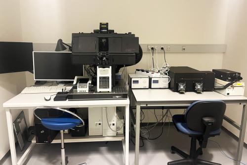 Olympus LSM FV1200S-IX83 confocal laser scanning microscope
Inverted - Laser Scanning Confocal Microscope with live cell incubator
FV1200 instruction video: https://video.ruc.dk/media/t/0_vi4kmkus
- Z-axis control for 3D imaging
- Motorized stage platform for tile-scan and multi-position imaging
- Time controller for time-lapse 4D imaging
- Differential Interference Contrast
- PECON on-stage incubator with controlled temperature, humidity and C02 for live cell/tissue/organoid imaging experiments
Detectors:
- 3 Photomultiplier tubes detectors (PMTs) (300 – 850 nm)
- 2 high-sensitivity GaAsp detectors (choice of YFP/GFP, Red/Near infrared or Red/green filter cubes). Can be used in combination with PMTs for multiplex imaging
- Transmitted light detector (T-PMT)
Laser lines:
- Laser diode 405 nm (DAPI, Photoactivation, Uncaging)
- Argon laser: 458, 488, 515 nm (CFP, GFP, YFP, Cy2, Alexa 488, FITC)
- DPSS Laser: 559 nm (RFPs, Cy3, Rhodamine Red-X, TRITC, Alexa 555/568)
- Red Diode Laser: 635 nm (Cy5, Alexa 633/647)
Objectives:
- UPLSAPO10X2 NA 0.40 WD 3.1 mm (air)
- UPLSAPO20X NA 0.75 WD 0.60 mm (air)
- UPLSAPO 30XS NA 1.05 WD 0.8 mm (silicone oil)
- UPLSAPO40X2 NA 0.95 WD 0.18 mm (air)
- UPLSAPO60XO NA 1.35 WD 0.15 mm (oil)
- UPLSAPO 100XS NA 1.35 WD 0.2 mm (silicone oil)
Fluorescence filter cubes:
- U-FUW (Wideband Ultraviolet excitation) – UV excitation: e.g. DAPI, Hoechst, Indo
- U-FBN (Narrowband Blue excitation) – Blue excitation: e.g. FITC, GFP/ EGFP, GFP2, Alexa Green, Fluo (Calcium indicator), Calcium green
- U-FGWA (Wideband Green excitation) – Green excitation: e.g. Rhodamine/TRITC, Cy3, Texas Red, Alexa Red, RFP
- U-F61001: Triple band filterset for DAPI/FITC/Propidium Iodid
-
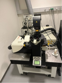 Leica TCS SP5 multi-photon microscope
Inverted Scanning Confocal Two-photon Microscope
- Two-photon (Multiphoton) imaging for deep tissue penetration and laser ablation experiments
- Z-axis control for 3D imaging
- Motorized stage platform for tile-scan and multi-position imaging
- Time controller for time-lapse 4D imaging
- Differential Interference Contrast
Laser lines:
- Argon 458/476/488/514nm
- DPSS 561nm
- HeNe 594nm
- HeNe 633nm
Detectors:
- Three photomultiplier tubes (PMTs) for fluorescence detection
- Two high-sensitivity HyD detectors
- Non-descanned Detector for two-photon
- Transmitted light detector (T-PMT)
Objectives:
- CS HC PL APO 10X NA 0.4 (air)
- HC PLAN APO 20X NA 0.7 (air)
- C5 HCX PL APO 63X NA 1.4 (oil)
- HCX PLAN APO 100X NA 1.35 (oil)
-
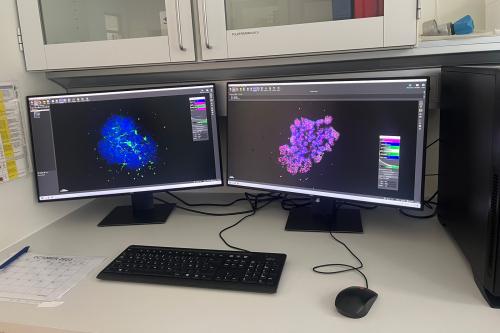 Image Analysis Workstation
Image analysis station and associated server drives for safe data storage
The computer has the following software installed:
- Continuously updated Imaris Full license (3D/4D image analysis software).
Includes Core module, Vantage, ClearView Deconvolution, Filament tracer, Cell analysis, Colocalization, Tracking, and Batch. - ImageJ/Fiji
- Ilastic
- CellProfiler
- Anaconda Navigator w Python and Jupyter Notebook
- Continuously updated Imaris Full license (3D/4D image analysis software).
-
We provide access to the following equipment for sample preparation, cell culture and basic microscopy in our laboratories:
- Cryostat
- Basic Stereomicroscopes
- Stereomicroscope equipped with a camera and fluorescence illumination (Zeiss)
- Widefield fluorescence microscopes
- GMO1 Cell laboratory with flow benches and incubators
- Fume hoods
- Freezers and refrigerators
- Various reagents for live cell imaging
-
We routinely maintain our microscopes, clean objectives, and perform quality controls using an Argolight calibration slide with power meter.


Activities
Courses
- RUC Molecular Health Master course: Experimental cell biology, runs every year in June and includes Confocal Microscopy elements
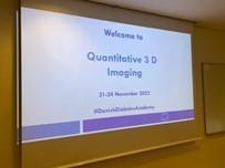
- Quantitative 3D imaging PhD course organized by Microscopy Core at RUC, DDEA & DBI-INFRA Image Analysis Core. Ran in 20222 and 2024 (https://ddeacademy.dk/events/course/quantitative-3d-bioimaging-phd-course/). Please enquire pianyeng@ruc.dk if interested in a future course.
- DBI-INFRA Image Analysis Core Facility courses: https://www.dbi-infra.eu/iacf-events
Events
- DBI events: https://www.danishbioimaging.dk/events/
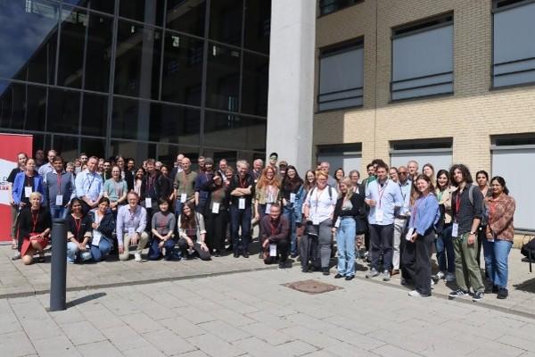
- The 8th Danish BioImaging Symposium was hosted at RUC in June 2025. The event focused on knowledge exchange and community building among bioimaging facilities and technology development groups.
- We participate in bi-monthly Danish Bioimaging Panel of core facilities (PoF) meetings
Booking, prices and preparation
-
To use the confocal microscopes, you need to book the system in advance.
- If you have not yet received training or if you are not staff at DSE, please write to microscope_core@ruc.dk, and arrange for training and booking.
- External users can only book our systems during work hours on weekdays.
- Members of staff at DSE who have received training may use the room booking system in Outlook:
• Leica SP5 TSC: Book Room 28AB.0-05
• Olympus FV1200: Book Room 28AB.0-07
• Imaris Image Analysis Workstation: Book Room 27A.1-29
- If you have not yet received training or if you are not staff at DSE, please write to microscope_core@ruc.dk, and arrange for training and booking.
-
Internal users and academic collaborators:
Projects running at DSE or related to DSE are free of charge.
Prices for visiting academia and commercial users:
Use of confocal microscopes with full support and/or training: 1335 DKK/hour
Use of confocal microscopes with minimal support: 813 DKK/hour
Use of confocal microscopes without support (after at least 2x training sessions): 639 DKK/hour
Personal support for sample preparation or image processing and analysis: 696 DKK/hour
Use of image analysis station, stereoscopes and laboratories are included free of charge for confocal microscopy users.
Prices are subject to change.Discounts:
When a booking exceeds 6 hours, subsequent hours, from 6 to 12 hours, are discounted 25%. Subsequent hours, following 12 hours usage, are discounted 50%.
-
Please contact us before initiating the experiment, to verify that your planned sample preparation is compatible with our systems.
Sample preparation checklist:
- Samples need to be mounted in appropriate medium with anti-fade reagents (e.g. the mounting media).
- Samples need to be covered with 0.17 mm thick coverslips. The coverslip should be sealed securely to the slide/dish holding the sample, as our systems are all inverted.
- In case of cell cultures, grow the cells on coverslips or in wells with 0.17 mm glass bottom. Keep in mind that all our confocal microscopes are inverted.
- Check compatibility of your fluorophores with our excitation laser lines (see Equipment)
- Samples need to be mounted in appropriate medium with anti-fade reagents (e.g. the mounting media).
-
Our Facility:
- Our FV1200 instruction video: https://video.ruc.dk/media/t/0_vi4kmkus
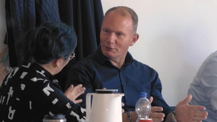
- Meet us and our users:
Our imaging community:
- DBI-INFRA: https://www.dbi-infra.eu/
- DBI Network: https://www.danishbioimaging.dk/
- Bridging Nordic Microscopy Infrastructure (BNMI): https://bnmi.eu/
- Euro-BioImaging: https://www.eurobioimaging.eu/
Resources
- FV1200 User guide: [link to PDF file]
- Leica TCS SP5: Product page
- iBiology – Fluorescence Microscopy Series: https://www.ibiology.org/online-biology-courses/microscopy-series/fluorescence-microscopy/
- Evident Scientific (OLYMPUS) – Confocal Microscopy Resources:
Tools and Calculators
- Nyquist Calculator: https://svi.nl/Nyquist-Calculator
- Fluorescent Protein Database & Spectra Viewer: https://www.fpbase.org/
Image Analysis
- ImageJ: Documentation | Forum
- Imaris Learning Resources: https://imaris.oxinst.com/learning/
-
For inquiries about using the confocal microscopes, please email microscope_core@ruc.dk. We are happy to assist with initial advice on experimental design, microscope choice and data analysis.
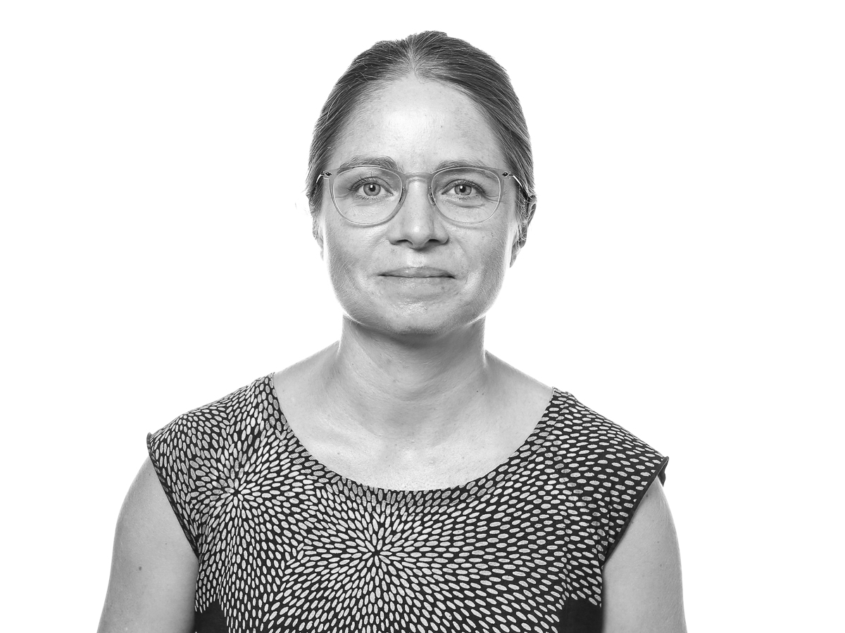 Pia Nyeng
Director
Pia Nyeng, Associate Professor and DBI-INFRA executive board member, pnyeng@ruc.dk
Expertise:
• Live organ explant and organoid imaging
• 3D multiplex imaging
• Quantitative image analysisStaff
• Christa Persson, laboratory technician
• Drisya Dileep, Postdoc
• Rikke Agerskov, PhD Student

Contact
Director
Pia Nyeng, Associate Professor
Phone: +45 4674 2883
Email: pnyeng@ruc.dk

Research facilities at Department of Science and Environment
Read more about the other research facilities at the Department of Sciences and Environment:
Research at the Department of Science and Environment - facilities